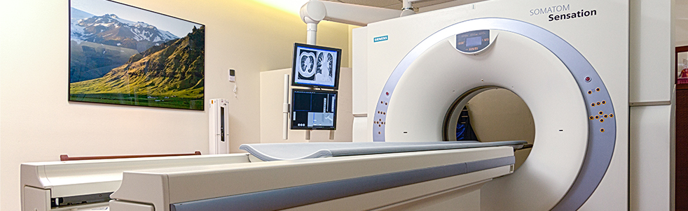


 X-ray, or radiography, is the most basic and common form of medical imaging.
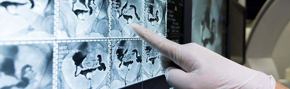
An X-ray machine produces a controlled beam of radiation, which is used to create an image of the inside of your body. This beam is directed at the area being examined. After passing through the body, the beam falls on a piece of film or a special plate where it casts a type of shadow. Different tissues in the body block or absorb the radiation differently. Dense tissue, such as bone, blocks most of the radiation and appears white on the film. Soft tissue, such as muscle, blocks less radiation and appears darker on the film. Often multiple images are taken from different angles so a more complete view of the area is available. The images obtained during X-ray exams are put through a process called “digitizing” so that they can be viewed on a computer screen.
Sometimes an X-ray exam includes contrast. For a contrast study, you will receive a drug called a contrast agent, which will highlight or contrast parts of the body so they show more clearly on the X-ray image.
X-ray exams can be used to view, monitor, or diagnose
- bone fractures
- joint injuries and infections
- artery blockages
- abdominal pain
- cancer
Preparation for an X-ray exam: For most X-ray exams, there is no special preparation needed. You will be asked to wear a hospital gown and remove all jewelry and metal objects before the test.
For contrast X-ray exams, you will be given a dose of contrast agent by mouth, as an enema, or as an injection or by catheter (thin tube) into a specific area of the body. Your physician will provide any specific instructions necessary for your contrast study.
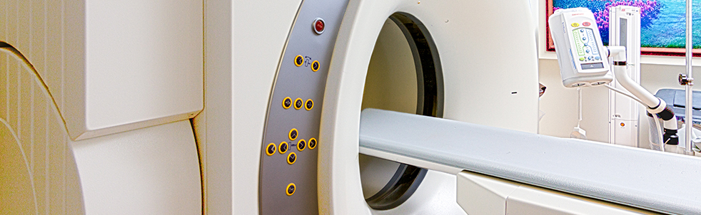
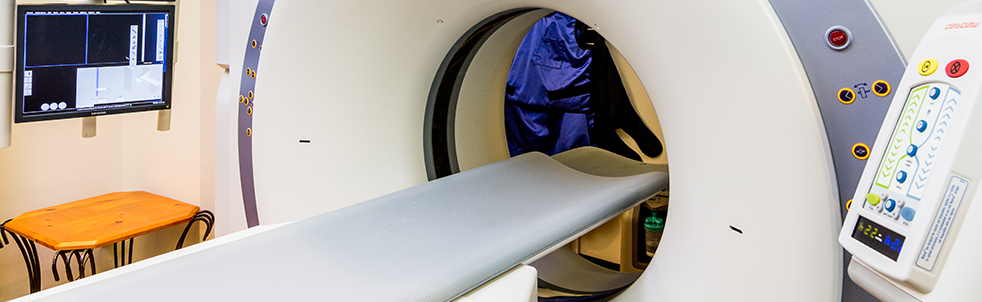
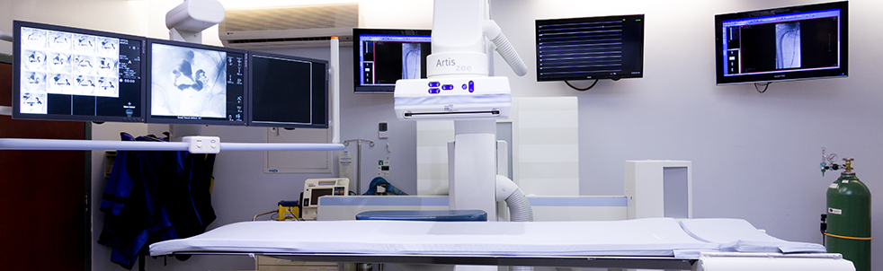
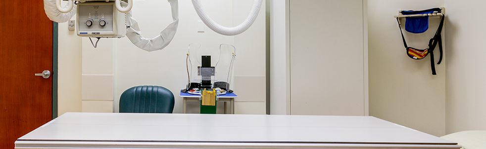
During the Exam: You will be asked to either lie on an exam table or stand next to the X-ray machine. The room may be cool in order to keep the equipment from overheating. The technologist, or person performing the exam, may use pillows or sandbags to help you hold the proper position. You will be asked to hold very still, without breathing for a few seconds. The technologist will step behind a radiation barrier and activate the X-ray machine. Often multiple images or views are taken from different angles, so the technologist will reposition you for another view and the process will be repeated. You will not feel the radiation.
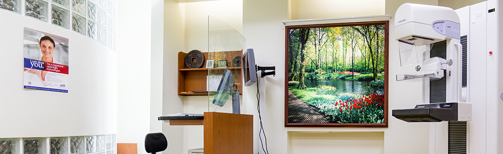
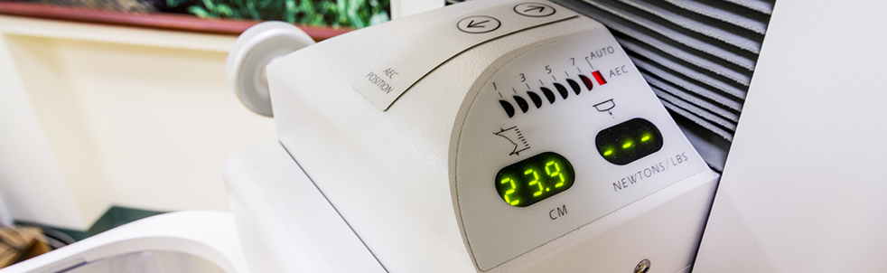
A mammogram is an X-ray exam of the breast. A special machine designed specifically to examine breast tissue is used. It takes a different form of X-ray and uses lower doses of radiation than a usual X-ray. Because these X-rays do not go through breast tissue as easily, the mammogram machine has two plates that compress the breast to spread the tissue apart. A more accurate image is obtained with less radiation this way.
Time Required: 5 to 60 minutes
Noise During Exam: Minimal clicking or buzzing noises.
Space During Exam: You will either lie on an exam table or stand next to the X-ray machine with ample space around you.
Benefits:
- X-ray exams are fast and easy.
- X-ray procedures are widely available.
Risks: X-ray exams exposure patients to radiation. The amount of radiation exposure is variable depending upon the X-ray type (for example, of the brain, lungs, or abdomen) and the X-ray machine type (for example, different models and manufacturers). Because the radiation exposure is variable, the risks are also variable. Please speak to your radiologist, or your physician who refers you for the X-ray exam, for specific details on radiation exposure and possible risks.
- Women should inform their doctor if they are or may be pregnant or nursing prior to any radiological imaging. Your doctor may recommend another type of test to reduce the possible risk of exposing your baby to radiation.
- There is a rare risk of a major allergic reaction to the contrast agent.
Results:
X-rays are recorded on film or recorded digitally. A radiologist, who is a physician with specialized training in X-ray and other imaging tests, will analyze and interpret the results of your X-ray and then send a report to your personal physician. For non-emergency situations, it usually takes a day or so to interpret, report, and deliver the results. Contact your personal physician for information on the results of your exam.
| Teleradiology | Women's Imaging Center |
|---|---|
|
At the GRC Women’s Imaging Center, we understand that women have unique healthcare concerns. We believe women deserve access to the specialized diagnostic services they need to prevent, detect and treat those ailments most common to them. GRC’s Women’s Imaging Center is designed to ensure that your specific health needs are addressed in a comprehensive and compassionate way. We have Guam’s most experienced women’s imaging team with Radiologists who have specialized in mammography as well as certified mammography, ultrasound and MRI technologists to ensure that your testing is accurate, comfortable and timely Read more... |

Contact Us
|
Guam Radiology Consultants |
Office Hours: Mon – Fri: 8AM – 6PM Sat: 8AM – 1PM |